Pet Retinal Detachments in Spring, TX
Get expert care for pet retinal detachments in Spring, TX, at North Houston Veterinary Ophthalmology. Our skilled specialists preserve your pet’s vision with timely treatment.

Retinal Detachments
Understanding Pet Retinal Detachments
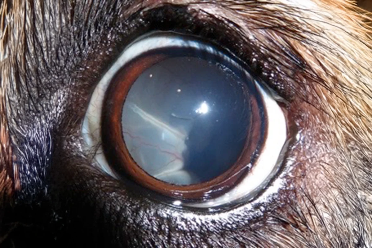
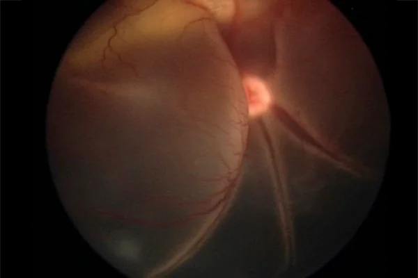
Retinal detachment in pets is a serious condition where the retinal layer separates from the choroid (the vascular nutritional layer) in the eye, leading to partial or complete blindness if not treated promptly. At North Houston Veterinary Ophthalmology, we are dedicated to diagnosing and treating retinal detachments to preserve your pet’s vision and maintain their quality of life.

Clinical Signs of Retinal Detachment
The clinical signs of retinal detachment vary, but common indicators include pupil dilation and sudden blindness. In cases where the retina is only partially detached, the loss of vision may be subtle and not immediately noticeable to pet owners. Other signs can include ocular redness, changes in the way your pet navigates familiar environments or bumping into objects.
Causes of Retinal Detachment
Retinal detachment can occur due to several underlying issues, including infectious or inflammatory diseases, high blood pressure, trauma, advanced cataracts, and chronic glaucoma. Our team at North Houston Veterinary Ophthalmology often encounters cases of retinal detachment stemming from genetic conditions such as progressive retinal atrophy, retinal dysplasia, merle ocular dysgenesis, and optic nerve or scleral colobomas. Some breeds, like Shih Tzus, may experience primary retinal detachments where no identifiable cause is present.
Treatment Options for Retinal Detachment
Prompt treatment of retinal detachment is crucial to prevent permanent vision loss. When the retina separates from the choroid, it suffers progressive damage that can lead to atrophy and complete loss of function. If the detachment is addressed within the first four weeks, there is a good chance that vision can be restored.
In many cases where retinal detachment is secondary to conditions like high blood pressure or infectious diseases, treating the underlying issue can allow the retina to reattach on its own. For partial detachments caused by trauma, surgery, or genetic conditions, laser treatment can be utilized to prevent further detachment and preserve existing vision. In severe cases where the retina has completely detached, referral for vitreo-retinal surgery might be necessary to reattach the retina and restore vision.
When to Seek Veterinary Ophthalmology Services
Pet owners should seek veterinary ophthalmology services if they notice any changes in their pet’s vision or eye appearance. Signs such as dilated pupils, sudden blindness, or noticeable changes in how your pet interacts with its environment indicate that immediate veterinary attention is needed. Early intervention can make a significant difference in the prognosis and treatment outcome for retinal detachment.
Comprehensive Care for Retinal Detachments
Retinal detachments are a serious condition that requires prompt and effective treatment to prevent permanent vision loss. North Houston Veterinary Ophthalmology is committed to providing comprehensive care for pets with retinal detachments. By understanding the clinical signs, causes, and treatment options, pet owners can make informed decisions and seek timely veterinary care for their beloved companions. Our team is here to ensure the best possible outcome for your pet’s vision and overall well-being.