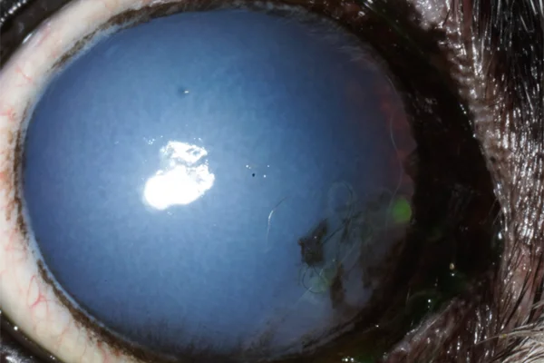
Pet Corneal Endothelial Degeneration in Spring, TX
Treat pet corneal endothelial degeneration at North Houston Veterinary Ophthalmology in Spring, TX, providing expert care and effective treatment to restore vision.

Pet Corneal Endothelial Degeneration
Understanding Pet Corneal Endothelial Degeneration
Corneal Endothelial Degeneration (CED) is a condition marked by fluid buildup (edema) in the cornea, leading to vision impairment. This condition primarily affects the cornea’s inner layer, the endothelium, and can significantly impact your pet’s quality of life. At North Houston Veterinary Ophthalmology, we specialize in diagnosing and treating corneal endothelial degeneration by providing comprehensive medical and surgical options.

Causes and Symptoms
What Causes Corneal Endothelial Degeneration?
CED is most common in certain breeds, such as Chihuahuas, Boston Terriers, and Dachshunds, but it can affect any breed. The condition arises from abnormal function of the corneal endothelium, which maintains corneal transparency by pumping out excess fluid. When this layer fails, fluid accumulates, leading to edema. Similar symptoms can also result from inflammation of the corneal endothelium.
Clinical Signs of Corneal Endothelial Degeneration
The primary clinical sign of CED is progressive corneal cloudiness, usually beginning laterally and spreading across the entire cornea over months to years. This cloudiness severely limits vision and can cause discomfort. In the later stages, pets may experience pain due to inflammation or corneal ulcers. It is essential to monitor your pet for these symptoms and seek veterinary care if you notice any changes in their eyes or behavior.
Diagnosing Corneal Endothelial Degeneration
North Houston Veterinary Ophthalmology employs a thorough diagnostic process to confirm the presence of CED. This involves a detailed examination of your pet’s eyes using specialized equipment to assess changes in the corneal layers.
Treatment Options for Corneal Endothelial Degeneration
In humans, CED is treated by transplanting the corneal endothelial layer. However, in pets, this is not currently available due to differences in pet eyes as compared to human eyes.
Surgical Grafting
The current most effective treatment for CED is a surgical grafting procedure called keratoleptynsis. This procedure involves advancing the scleral conjunctiva over the affected portions of the cornea to decrease and slow the progression of corneal edema. The goal is to restore vision and alleviate discomfort by reducing fluid buildup.
Importance of Early Intervention
The best results are achieved when surgery is performed in the earlier stages before significant vision loss occurs. Early intervention offers the best chance of preventing progression of this condition and improves the overall prognosis of preserving vision and preventing painful recurrent corneal ulceration. It is crucial to seek veterinary care as soon as you notice any signs of corneal cloudiness or vision impairment in your pet.
Postoperative Care
Postoperative care is vital for a successful recovery. Your veterinary ophthalmologist will provide detailed instructions on administering medications, such as anti-inflammatory drugs and antibiotics, to prevent infection and reduce swelling. Regular follow-up visits are necessary to monitor your pet’s progress and address any complications.
Benefits of Treating Corneal Endothelial Degeneration
Treating CED helps to maintain vision, allowing your pet to navigate their environment comfortably. Early treatment alleviates discomfort from corneal edema and prevents painful corneal ulcers. Addressing CED early enhances your pet’s quality of life, ensuring they remain active and happy.
When to Seek Veterinary Care
Seek veterinary care as soon as you notice any signs of CED. Early intervention can prevent the condition from worsening and reduce the risk of complications. If you observe progressive corneal cloudiness, vision impairment, or signs of discomfort, contact North Houston Veterinary Ophthalmology in Spring, TX, immediately. Our team provides compassionate and comprehensive care to ensure your pet’s eyes remain healthy.
Expert Care for Pet Corneal Endothelial Degeneration
Pet corneal endothelial degeneration is a serious condition requiring prompt and professional treatment. At North Houston Veterinary Ophthalmology in Spring, TX, we are committed to providing the highest standard of care. Understanding the causes, symptoms, and treatment options for this condition ensures your pet receives the best possible care. If you suspect your pet has corneal endothelial degeneration, reach out to our clinic. Our committed staff is here to assist in achieving ideal eye health and preserving your pet’s excellent standard of living.